E coli under microscope 4x 232005
It is estimated that % of the human population are longterm carriers of S aureusE) electron microscope compound light microscope 10 x 4x= 40 (TM) What is the total magnification for the low lens 10 x 10x= 100 (TM) What is the total magnification for the high lens 10 x 40x= 400 (TM) e coli What is an Acid Fast cells that retain a basic stain in the presence of an acidalcoholThe virulent E coli bacterium that killed dozens of people will shortly land under the microscope as part of a European project to better understand dangerous pathogens
Q Tbn And9gcrxsv6valktvydnaiqm6 Yoim4dbn 9td0atqk2azvfczn73wcq Usqp Cau
E coli under microscope 4x
E coli under microscope 4x-479 KB Escherichia coli flagella TEMpng 600 × 6;Immersion oil onto the part of the slide directly under the lens Now, using the fine adjuster ONLY, focus in onto your organism Those of you using E coli will see something like this
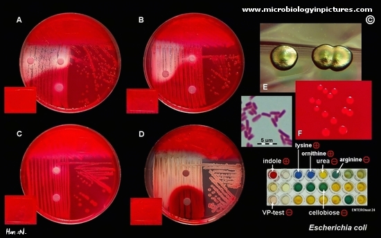


Escherichia Coli Colony Morphology And Microscopic Appearance Basic Characteristic And Tests For Identification Of E Coli Bacteria Images Of Escherichia Coli Antibiotic Treatment Of E Coli Infections
Lens at the top of microscope 🔬 that users lookWhat will you be able to see under a high power microscope?First, by using Escherichia coli (Ecoli), we start at 4x objective, followed by low power 10x objective viewing and the specimen looked clearer at 40x magnificationThe shaped of Ecoli is rodshaped and it is a gramnegative bacteriaThe gram stain of some specimen either gramnegative or grampositive can be resulted by looked at their color whether the bacteria are pink or red for gram
Cheek Cells Under a Microscope Requirements, Preparation and Staining Cheek cells are eukaryotic cells (cells that contain a nucleus and other organelles within enclosed in a membrane) that are easily shed from the mouth lining It's therefore easy to obtain them for observationFurther, a keen observation under the microscope clearly indicated that, C elegans coculture with A courvalinii JP_A1001, significantly (p ** < 001) affected their body bends/s when compared to E coli OP50 (Fig 6D)No special preparation of fixation or staining etc is needed to study an object under a phasecontrast microscope which saves a lot of time Examining intracellular components of living cells at relatively high resolution eg The dynamic motility of mitochondria , mitotic chromosomes & vacuoles
A rectangular piece of glass where a sample is mounted for viewing under the microscope What is the eye piece?Mount a piece of a Coleus leaf on a microscope slide Be sure to have the 4X objective in place Use tape to secure the leaf at each end Do NOT cover the leaf with tape!!!263 MB Escherichia coli electron microscopyjpg 800 × 640;


Lab 1


What Does An E Coli Bacteria Look Like Under A Microscope Quora
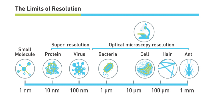
The microscopes I use in my bacteriology lab have a 10x eyepiece and a selection of objective lenses with the following magnifications 1 4x 2 10x 3 40x 4 100x With the magnification of the eyepiece, these objectives give magnifications of 4Yes, most of the bacteria range from 022 µm in diameter The length can range from 110 µm for filamentous or rodshaped bacteria The most wellknown bacteria E coli, their average size is ~15 µm in diameter and 26 µm in length As we talked above, you can see some bacteria in my cheek cellsIt is purposely done that way so that the lab technician can easily differentiate the cells when viewing under the microscope Under the microscope, the vegetative cells appear reddish to pinkish in color while the endospores are green in color when examined under the microscope (9, 10) #2 – Dorner's Method


Aph162 Report 1


Escherichia Coli Responds To Environmental Changes Using Enolasic Degradosomes And Stabilized Dicf Srna To Alter Cellular Morphology Pnas
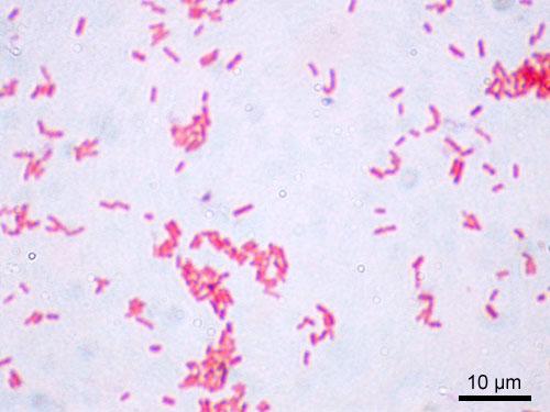
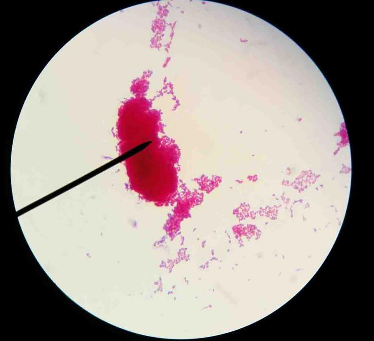
Examples of Bacteria Under the Microscope Escherichia coli Escherichia coli (Ecoli) is a common gramnegative bacterial species that is often one of the first ones to be observed by students Most strains of Ecoli are harmless to humans, but some are pathogens and are responsible for gastrointestinal infections They are a bacillus shapedFigure 2 Under the microscope, E coli was stained reddish pink, which means it is Gram negative It was hard to observe the morphological structure of the bacteria, because it was very clustered The slide was observed at 4x also DISCCUSION During the observation, it was hard to see each bacterium because they were too clustered To prevent this, it is important to obtain small amount ofFigure 2 Under the microscope, E coli was stained reddish pink, which means it is Gram negative It was hard to observe the morphological structure of the bacteria, because it was very clustered The slide was observed at 4x also DISCCUSION During the observation, it was hard to see each bacterium because they were too clustered To prevent this, it is important to obtain small amount of


Escherichia Coli Colony Morphology And Microscopic Appearance Basic Characteristic And Tests For Identification Of E Coli Bacteria Images Of Escherichia Coli Antibiotic Treatment Of E Coli Infections


Observing Bacteria Under The Light Microscope Microbehunter Microscopy
Approximately 2lum Organelles in ecoli Cell wall, cell membrane,cytoplasm,flagellum,pilus,nucleoid(dna) 4x,10x,40x What is the slide?The prepared microscope slide image of Bacillus Subtilis at left was captured at 400x magnification Learn more here E Coli E Coli under the microscope at 400x E Coli (Escherichia Coli) is a gramnegative, rodshaped bacterium Most E Coli strains are harmless, but some serotypes can cause food poisoning in their hostsResearcher with e coli bacteria bacteria under microscope stock pictures, royaltyfree photos & images ebola outbreak infographic bacteria under microscope stock illustrations chemical test of water bacteria under microscope stock pictures, royaltyfree photos & images
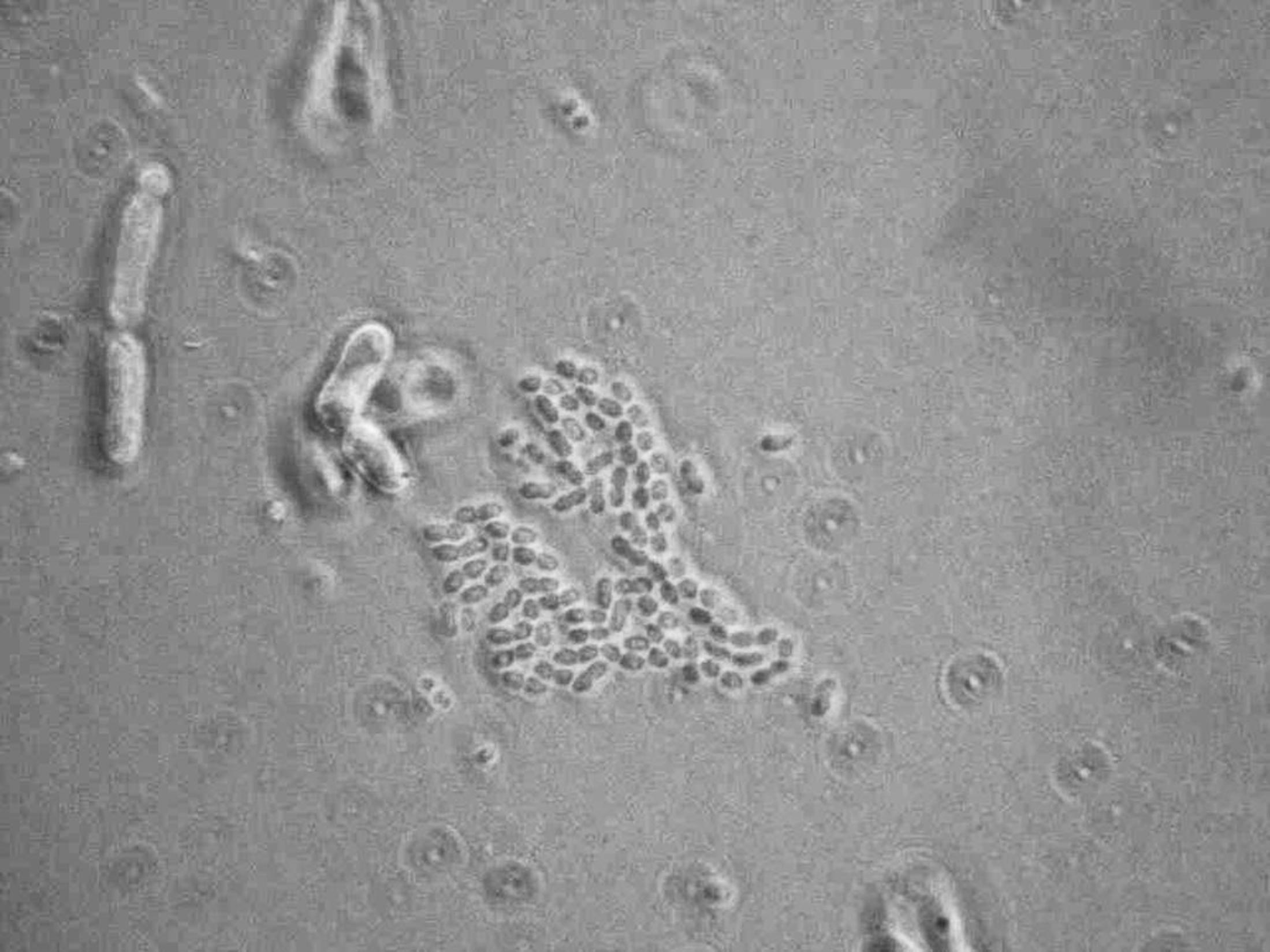


Microscopy For The Winery Viticulture And Enology


What Does An E Coli Bacteria Look Like Under A Microscope Quora
Escherichia coli and Staph aureus ()jpg 2,160 × 1,440;E Coli Under The Microscope Types Techniques Gram Stain Escherichia Coli E Coli Microscope Bacterial Shape Coccus 1000x General Biology Lab Loyola Microscopy For The Winery Viticulture And Enology Onion Cell Under Microscope 4x 10x 40xKlebsiella ranks second to E coli for urinary tract infections in older persons It is also an opportunistic pathogen for patients with chronic pulmonary disease, enteric pathogenicity, nasal mucosa atrophy, and rhinoscleroma Feces are the most significant source of patient infection, followed by contact with contaminated instruments


Rose Gold Collection 3 Modern Stock Footage Video 100 Royalty Free Shutterstock


Biol 230 Lab Manual Lab 1
LAB 3 – Use of the Microscope Introduction 4X – This objective magnifies the image by a factor of 4 Get a slide of the letter "e" from the tray on the side counter This an example of a prepared slide, a slide that is already made for you and meant to be reusedPlace the slide on the microscope stage Move it so that the leaf is under the objective Be sure that the condenser iris is wide openWhen viewed under the microscope, Gramnegative E Coli will appear pink in color The absence of this (of purple color) is indicative of Grampositive bacteria and the absence of Gramnegative E Coli Escherichia coli under 10х90х magnification using fuchsine as a dye by ElNokko (Own work) CC BYSA 40 (http//creativecommonsorg/licenses/bysa/40), via Wikimedia Commons


Gram Stain
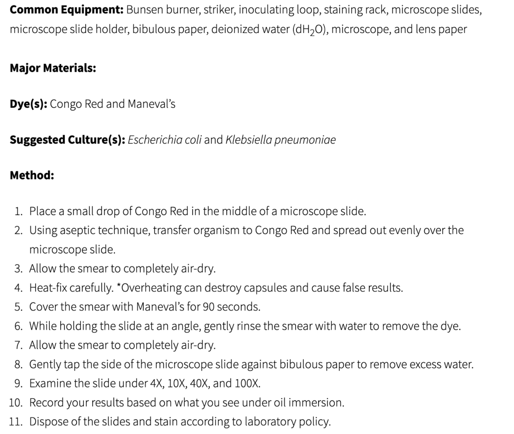


Solved Need Help With The Blank Spaces Below Capsule St Chegg Com
Escherichia coli bacteria on blood agar e coli under a microscope stock pictures, royaltyfree photos & images petri dishes with culture media for sarscov2 diagnostics, test coronavirus covid19, microbiological analysis e coli under a microscope stock pictures, royaltyfree photos & imagesWhat is the approximate size of Ecoli?Cheek Cells Under a Microscope Requirements, Preparation and Staining Cheek cells are eukaryotic cells (cells that contain a nucleus and other organelles within enclosed in a membrane) that are easily shed from the mouth lining It's therefore easy to obtain them for observation


Http Coltonanderson1 Weebly Com Uploads 2 4 3 0 Manual Pdf


Time Kill Curves Gram Negative Strains P Aeruginosa A And E Coli Download Scientific Diagram
Researcher with e coli bacteria bacteria under microscope stock pictures, royaltyfree photos & images ebola outbreak infographic bacteria under microscope stock illustrations chemical test of water bacteria under microscope stock pictures, royaltyfree photos & imagesE coli is one of the key prokaryotic model organisms used in the fields of biotechnology and microbiology Hence, in many recombinant DNA experiments, E coli serves as the host organism The reasons behind using E coli as the primary model organism are some characteristics of E coli such as fast growth, availability of cheap culture media to grow, easiness to manipulate, extensiveEnsure the microscope is on the lowest magnification The part of the microscope that allows for magnification is called an objective Objectives for compound light microscopes usually range from 4x to 40x You can move to higher magnifications if necessary, but starting low allows you to easily find the specimen on the slide


Flower Like Patterns In Multi Species Bacterial Colonies Elife


Lab 1
Microorganisms are important Recent news topics have focused on a variety of microorganisms E coli K12 in food contamination, life on Mars, Ebola virus in Africa, AIDS, and the recent rise in Tuberculosis infections Size is an important characteristic that students might investigate as they attempt to put these organisms into perspectiveE coli under microscope By Barbara Duckworth Reading Time 3 minutes Published October 12, 00 Livestock The quest to unravel the mystery of E coli bacteria grows more perplexing4 Can a light microscope see bacteria?


What Does An E Coli Bacteria Look Like Under A Microscope Quora


New Flat Embedding Method For Transmission Electron Microscopy Reveals An Unknown Mechanism Of Tetracycline Biorxiv
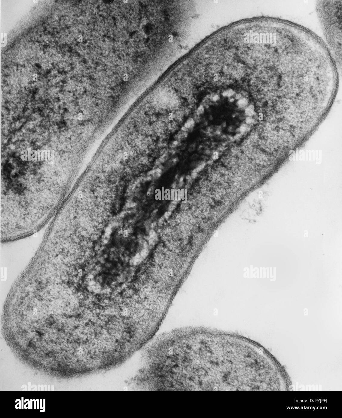
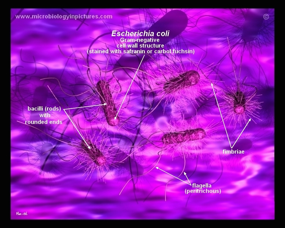
E coli under the microscope Matthew Tessnear Tuesday Oct 16, 12 at 11 AM Oct 16, 12 at 525 PM Health officials say they expect a slow down soon in the number of reported new E coli cases in an outbreak linked to the Cleveland County FairEscherichia coli (/ ˌ ɛ ʃ ə ˈ r ɪ k i ə ˈ k oʊ l aɪ /), also known as E coli (/ ˌ iː ˈ k oʊ l aɪ /), is a Gramnegative, facultative anaerobic, rodshaped, coliform bacterium of the genus Escherichia that is commonly found in the lower intestine of warmblooded organisms (endotherms) Most E coli strains are harmless, but some serotypes (EPEC, ETEC etc) can cause serious foodThe nucleoid of living and OsO4 or glutaraldehydefixed cells of Escherichia coli strains was studied with a phasecontrast microscope, a confocal scanning light microscope, and an electron microscope The trustworthiness of the images obtained with the confocal scanning light microscope was investigated by comparison with phasecontrast micrographs and reconstructions based on serially


Q Tbn And9gcrxsv6valktvydnaiqm6 Yoim4dbn 9td0atqk2azvfczn73wcq Usqp Cau


Lab Manual Exercise 1
Recent news topics have focused on a variety of microorganisms E coli K12 in food contamination, life on Mars, Ebola virus in Africa, AIDS, and the recent rise in Tuberculosis infections Size is an important characteristic that students might investigate as they attempt to put these organisms into perspectiveExamples of Bacteria Under the Microscope Escherichia coli Escherichia coli (Ecoli) is a common gramnegative bacterial species that is often one of the first ones to be observed by students Most strains of Ecoli are harmless to humans, but some are pathogens and are responsible for gastrointestinal infections They are a bacillus shaped bacteria that has a very fast growth (they can double every minutes), which is one of the main reasons they are used in researchSep 19, 13 Explore Julie Marsh's board "E coli", followed by 181 people on See more ideas about microbiology, microscopic photography, ecoli


Schematic Of E Coli Bioenergetics With High Energy Compounds Atp And Download Scientific Diagram


Figures And Data In Flower Like Patterns In Multi Species Bacterial Colonies Elife
Yes, most of the bacteria range from 022 µm in diameter The length can range from 110 µm for filamentous or rodshaped bacteria The most wellknown bacteria E coli, their average size is ~15 µm in diameter and 26 µm in length As we talked above, you can see some bacteria in my cheek cellsE coli is a Gramnegative rodshaped bacteria When Gram stained, the organism looks pink or red Here are a couple of pictures of a Gram stain of E coli that I did under the 100X objective lens on a standard light microscope You can of courseMicroscope World recently took a Gram stain prepared slide of three different types of bacteria and captured images using different microscope objective lenses The microscope used to view the bacteria was the Fein Optic R0 biological lab microscope
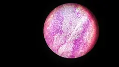


E Coli Under The Microscope Types Techniques Gram Stain Hanging Drop Method


Microscope World Blog Bacillus Subtilis Under The Microscope
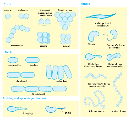
When using a high power microscope (also known as a compound microscope) it is best to start out with the lowest magnification, get your specimen in focus, and then move up to the higher magnifications one at a time This is the easiest way to ensure that you will be able to focus inE coli under the microscope Escherichia coli (E coli) is a bacterium commonly found in various ecosystems like land and water Most of the strains of E coli are harmless, but some strains are known to cause diarrhea and even UTIs E coli is commonly studied as they are considered as a standard for the study of different bacteriaCocci in grapelike clusters (Saureus) and bacilli(Ecoli) Clinical significance of Saureus Frequently found as part of the normal skin flora on the skin and nasal passages;


Yoghurt Bacteria Experiments On Microscopes 4 Schools


Gut Bacteria Escherichia Coli Under Microscope Youtube
E coli is one of the key prokaryotic model organisms used in the fields of biotechnology and microbiology Hence, in many recombinant DNA experiments, E coli serves as the host organism The reasons behind using E coli as the primary model organism are some characteristics of E coli such as fast growth, availability of cheap culture media to grow, easiness to manipulate, extensive4 Can a light microscope see bacteria?E Coli E Coli under the microscope at 400x E Coli (Escherichia Coli) is a gramnegative, rodshaped bacterium Most E Coli strains are harmless, but some serotypes can cause food poisoning in their hosts The harmless strains are part of the normal flora of the gut Learn more about E Coli here Helicobacter Pylori


Microscope World Blog June 15


Aph162 Report 1
1 Turn on microscope, firstly use 4x lenses and then up to 40x to magnify image of specimen 2 take clean slides and label them as following BSubtilis àCV Pvulgaris àHDT (use depression slide) Saureus àMB ScerevisiaeàTWM Ecoli àS First we cleaned the objectives of the microscope with cleaning solution and cleaned the benches withResearcher with e coli bacteria bacteria under microscope stock pictures, royaltyfree photos & images ebola outbreak infographic bacteria under microscope stock illustrations chemical test of water bacteria under microscope stock pictures, royaltyfree photos & imagesE Coli Under The Microscope Types Techniques Gram Stain Escherichia Coli E Coli Microscope Bacterial Shape Coccus 1000x General Biology Lab Loyola Microscopy For The Winery Viticulture And Enology Onion Cell Under Microscope 4x 10x 40x


Methods In Cell Biology


Laboratory Test 1 Flashcards Chegg Com
The easiest way to see bacteria through a microscope is to prepare a bacterial smear A smear is a bacterial sample mixed into a drop of water on a microscope slide and then heat fixed By placing the slide on a microincinerator tray or gently waving slide over Bunsen burner flame until dryGet a second escherichia coli bacteria (e coli) stock footage at 2997fps 4K and HD video ready for any NLE immediately Choose from a wide range of similar scenes Video clip id Download footage now!The prepared microscope slide image of Bacillus Subtilis at left was captured at 400x magnification Learn more here E Coli E Coli under the microscope at 400x E Coli (Escherichia Coli) is a gramnegative, rodshaped bacterium Most E Coli strains are harmless, but some serotypes can cause food poisoning in their hosts


Bacteria Micrograph High Resolution Stock Photography And Images Alamy


New Flat Embedding Method For Transmission Electron Microscopy Reveals An Unknown Mechanism Of Tetracycline Biorxiv


Solved 1 Which Would Be The Greatest Potential Source Of Chegg Com


How E Coli Bacteria Look Like
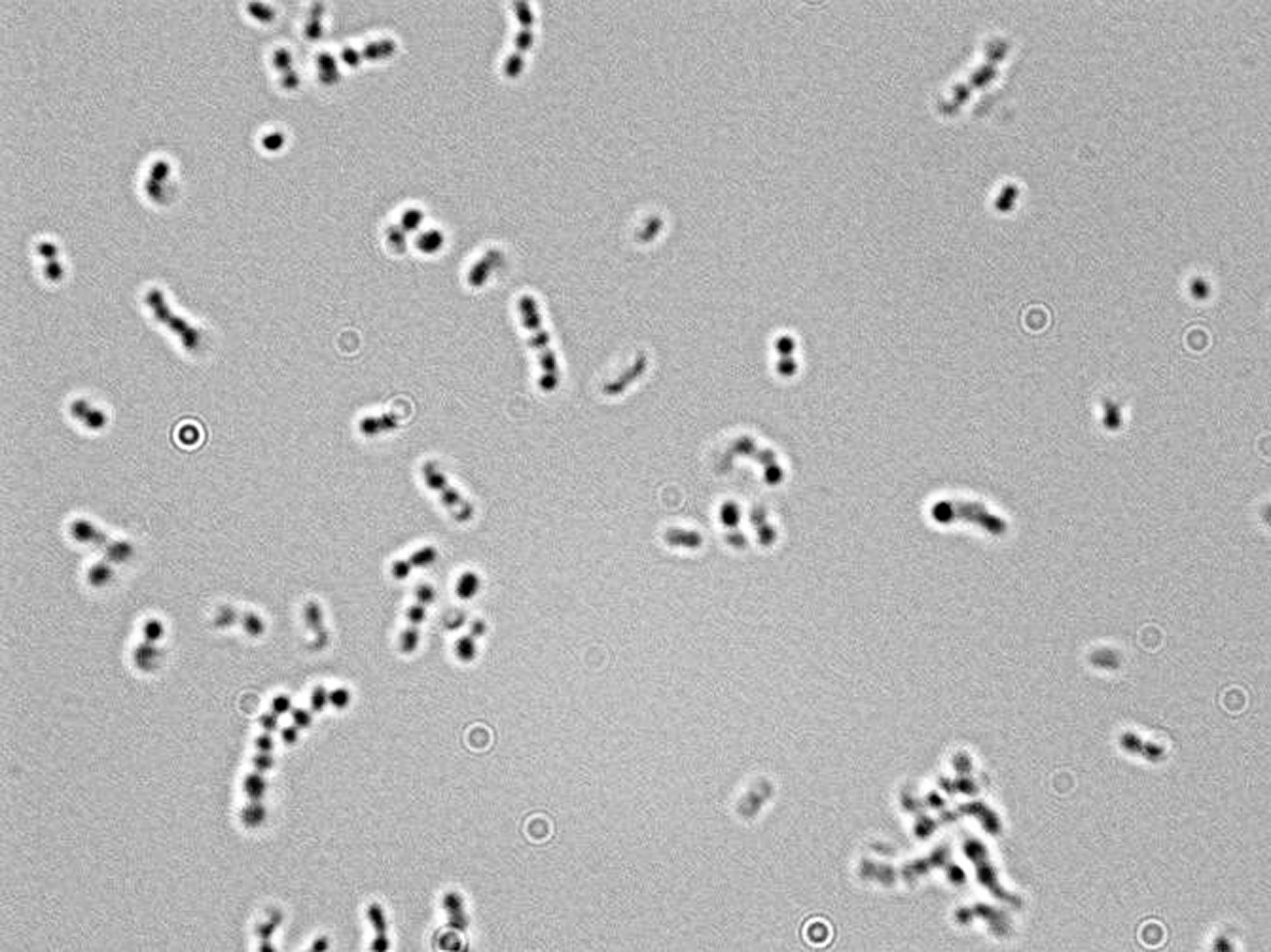


Microscopy For The Winery Viticulture And Enology


1b Demo


Gram Stain


Zkfaa Bioproses Lab 1 Principles And Use Of Microscope


Staphylococcus Aureus And Ecoli Under Microscope Microscopy Of Gram Positive Cocci And Gram Negative Bacilli Morphology And Microscopic Appearance Of Staphylococcus Aureus And E Coli S Aureus Gram Stain And Colony Morphology On Agar Clinical


Escherichia Coli Responds To Environmental Changes Using Enolasic Degradosomes And Stabilized Dicf Srna To Alter Cellular Morphology Pnas


Staphylococcus Aureus And Ecoli Under Microscope Microscopy Of Gram Positive Cocci And Gram Negative Bacilli Morphology And Microscopic Appearance Of Staphylococcus Aureus And E Coli S Aureus Gram Stain And Colony Morphology On Agar Clinical


Escherichia Coli Colony Morphology And Microscopic Appearance Basic Characteristic And Tests For Identification Of E Coli Bacteria Images Of Escherichia Coli Antibiotic Treatment Of E Coli Infections
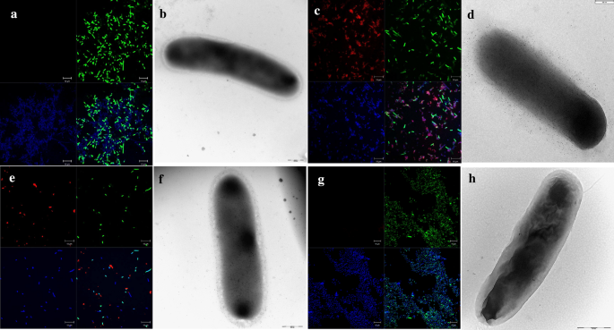


Decorating The Surface Of Escherichia Coli With Bacterial Lipoproteins A Comparative Analysis Of Different Display Systems Microbial Cell Factories Full Text
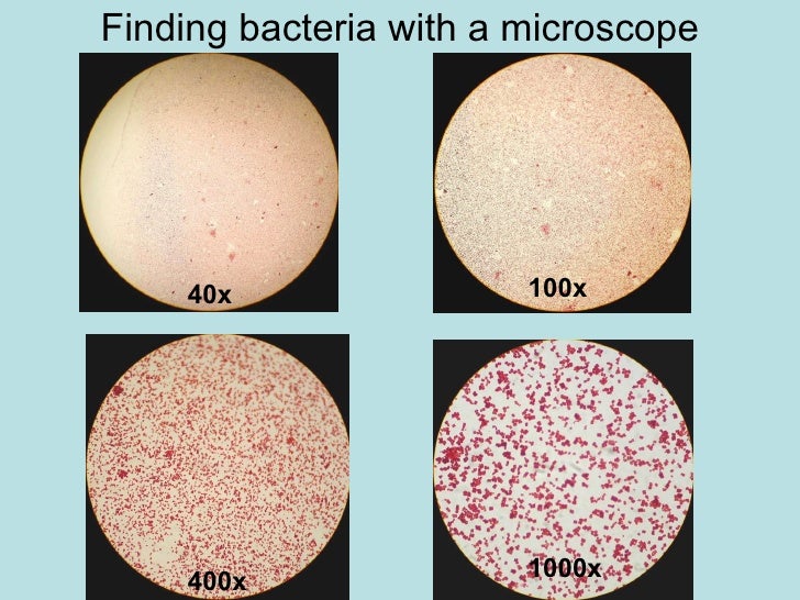


Chapter 3 Tools Of The Laboratory E Mail


Observing Bacteria Under The Light Microscope Microbehunter Microscopy


Flower Like Patterns In Multi Species Bacterial Colonies Elife


Q Tbn And9gcqkye60ou Johpr02n Mbv1fferrjpdh Lnct7ymdf5qhyia1ld Usqp Cau


Phagocytosis Assay Kit Red E Coli Bn Assay Genie


Bacteria Under The Microscope Microscope And Laboratory Equipment Reviews


Gram Stain Kit Advanced With Stabilized Iodine Produces Brighter Colors For Both Gram Positives And Negatives 4 X 8oz Bottles Emerald Scientific
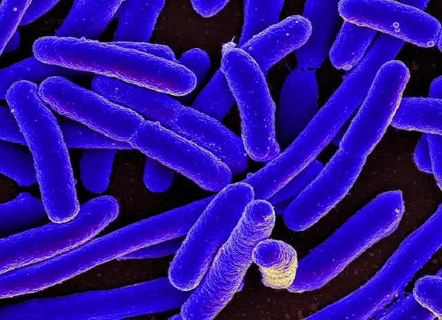


E Coli Under The Microscope Types Techniques Gram Stain Hanging Drop Method


Escherichia Coli Light Microscopy
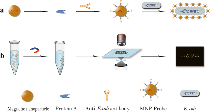


Ultrasensitive And Rapid Count Of Escherichia Coli Using Magnetic Nanoparticle Probe Under Dark Field Microscope Bmc Microbiology Full Text


Escherichia Coli Bacteria E Coli Stock Footage Video 100 Royalty Free Shutterstock


Observing Bacteria Under The Light Microscope Microbehunter Microscopy
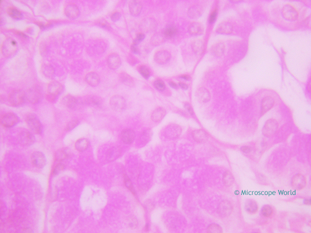


Why Would You Need Microscope Immersion Oil And How To Use It


Escherichia Coli Bacteria E Coli Stock Footage Video 100 Royalty Free Shutterstock


Observing Bacteria Under The Light Microscope Microbehunter Microscopy


Escherichia Coli Bacteria E Coli Stock Footage Video 100 Royalty Free Shutterstock


10pk Escherichia Coli Smear Gram Stain Prepared Microscope Slides 75 X 25mm Classroom Pack 10 Slides In Storage Case Biology Microscopy Eisco Labs Amazon Com Industrial Scientific


New Flat Embedding Method For Transmission Electron Microscopy Reveals An Unknown Mechanism Of Tetracycline Biorxiv


Flower Like Patterns In Multi Species Bacterial Colonies Elife


What Does An E Coli Bacteria Look Like Under A Microscope Quora


Pseudomonas Aeruginosa Promotes Escherichia Coli Biofilm Formation In Nutrient Limited Medium


What Does An E Coli Bacteria Look Like Under A Microscope Quora
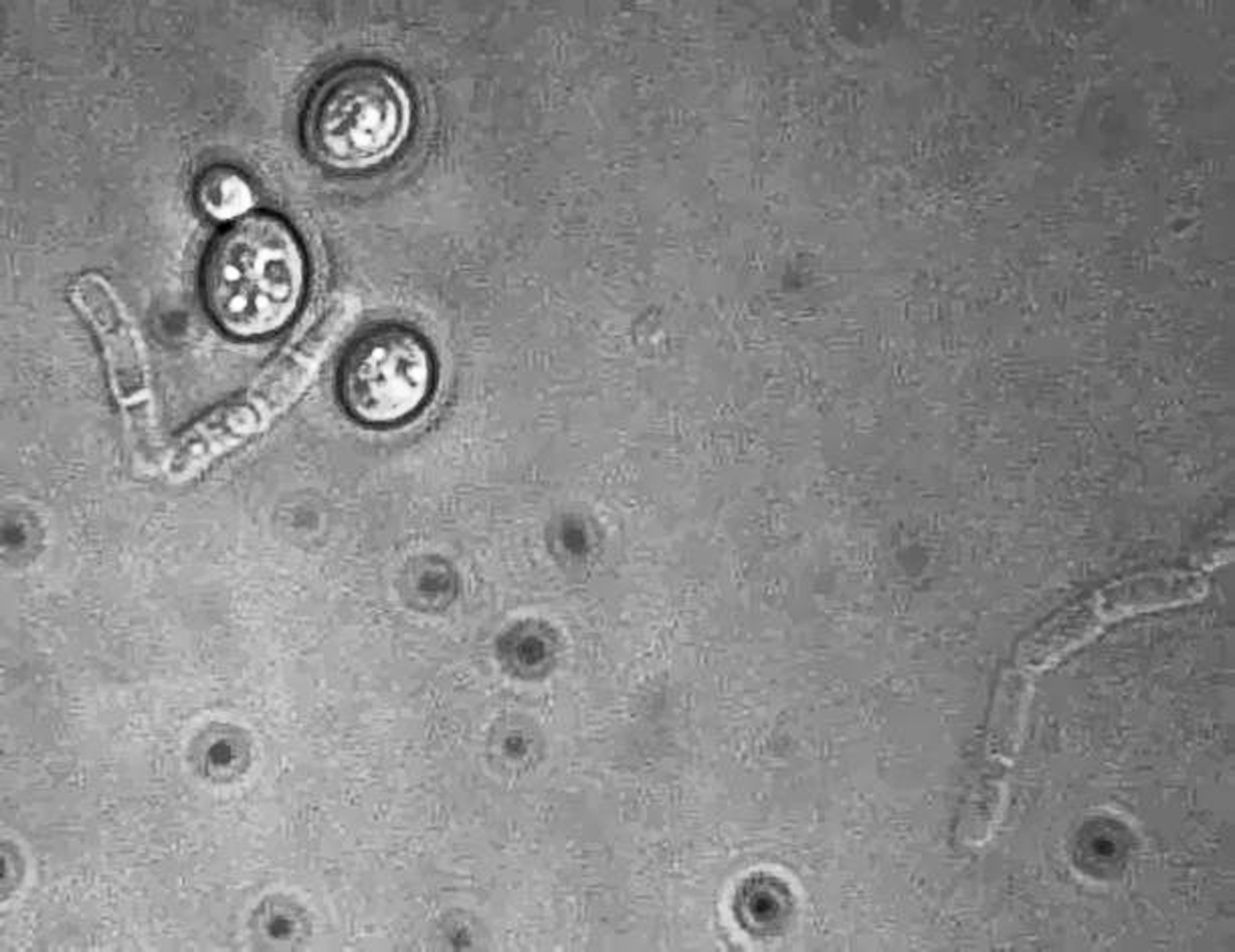


Microscopy For The Winery Viticulture And Enology


What Does An E Coli Bacteria Look Like Under A Microscope Quora


Terrestrial Landscape Impacts The Biogeographic Pattern Of Soil Escherichia Coli Via Altering The Strength Of Environmental Selection And Dispersal Biorxiv


Team Leicester August12 12 Igem Org


Gut Bacteria Escherichia Coli Under Microscope Youtube


Gram Stain


Escherichia Coli Bacteria E Coli Stock Footage Video 100 Royalty Free Shutterstock


Zkfaa Bioproses Lab 1 Principles And Use Of Microscope


Microscope World Blog June 15
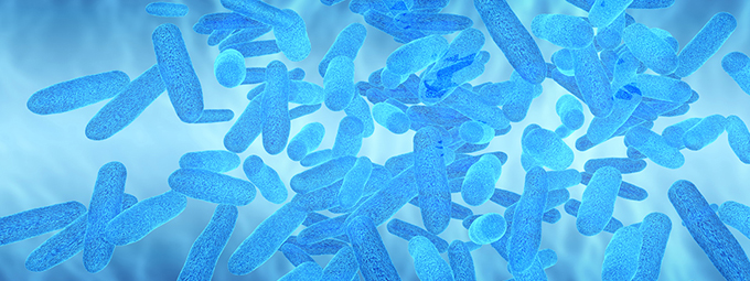


What Magnification Do I Need To See Bacteria Westlab
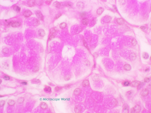


Why Would You Need Microscope Immersion Oil And How To Use It


Microscope World Blog June 15


Whats Your Microscopy Setup Advanced Mycology Shroomery Message Board


Lab 1
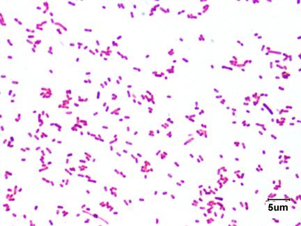


Laboratory Test 1 Flashcards Chegg Com
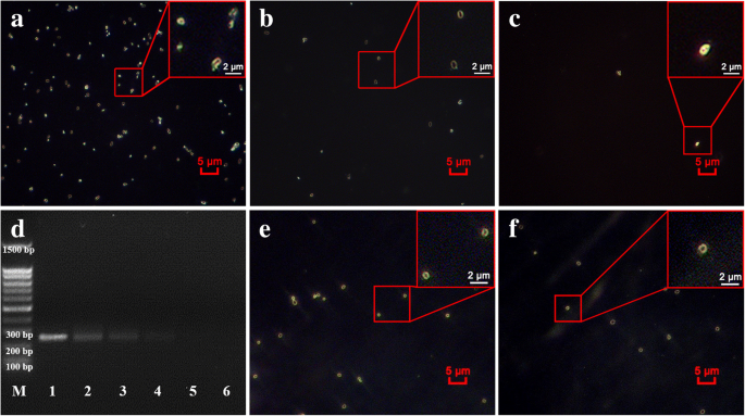


Ultrasensitive And Rapid Count Of Escherichia Coli Using Magnetic Nanoparticle Probe Under Dark Field Microscope Bmc Microbiology Full Text


9 Questions With Answers In Escherichia Coli Science Topic


Observing Bacteria Under The Light Microscope Microbehunter Microscopy
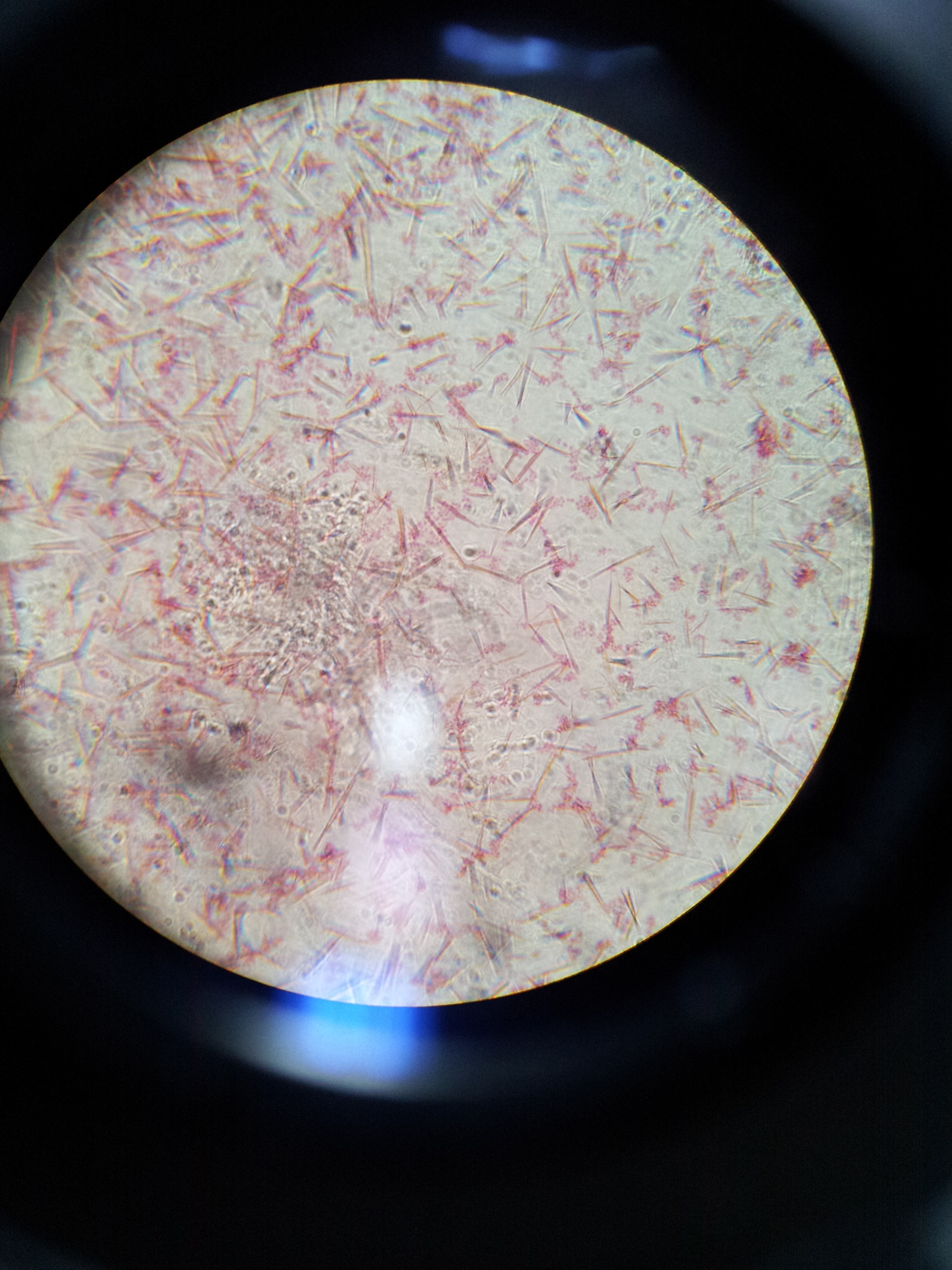


Lab 1 Principles And Use Of Microscope Ibg 102 Lab Reports


Pseudomonas Aeruginosa Promotes Escherichia Coli Biofilm Formation In Nutrient Limited Medium


Micro Lab Final Flashcards Quizlet


Lab 1 Principles And Use Of Microscope


Staphylococcus Aureus And Ecoli Under Microscope Microscopy Of Gram Positive Cocci And Gram Negative Bacilli Morphology And Microscopic Appearance Of Staphylococcus Aureus And E Coli S Aureus Gram Stain And Colony Morphology On Agar Clinical


Biol 230 Lab Manual Lab 1


What Magnification Do I Need To See Bacteria Westlab


Q Tbn And9gcr1gxralqmge Lwsjjzsouihvmyiv0ajl7a5fu8ma5voxpjs64l Usqp Cau


Acid Fast Stain Free Microbiology Images From Science Prof Online


E Coli Escherichia Coli A Gram Negative Bacterium Causing Gastrointestinal Infection


Observing Bacteria Under The Light Microscope Microbehunter Microscopy
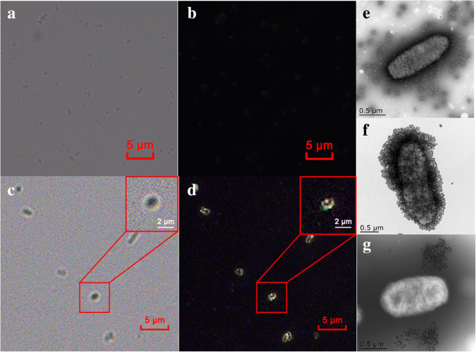


Ultrasensitive And Rapid Count Of Escherichia Coli Using Magnetic Nanoparticle Probe Under Dark Field Microscope Bmc Microbiology Full Text


Q Tbn And9gcqkye60ou Johpr02n Mbv1fferrjpdh Lnct7ymdf5qhyia1ld Usqp Cau


Bacteria Under The Microscope E Coli And S Aureus Youtube


Microscope World Blog June 15


10pk Escherichia Coli Smear Gram Stain Prepared Microscope Slides 75 X 25mm Classroom Pack 10 Slides In Storage Case Biology Microscopy Eisco Labs Amazon Com Industrial Scientific


Gram Stain
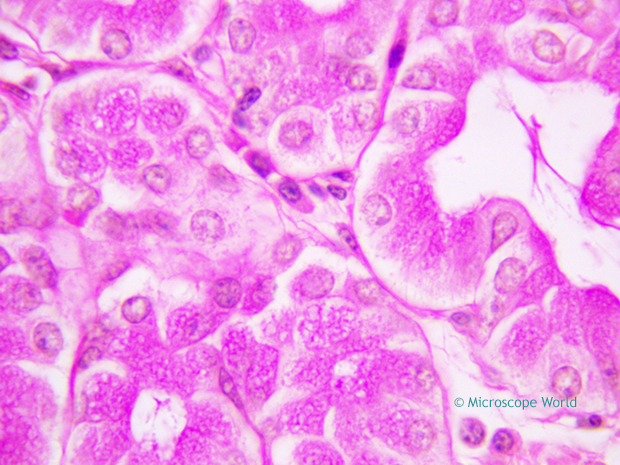


Why Would You Need Microscope Immersion Oil And How To Use It


Entamoeba Coli Best View In Stool Microscopy At 40x E Coli In Stool Microscopy Parasites In Stool Youtube


Lab 1 Principles And Use Of Microscope


Biol 230 Lab Manual Lab 1


コメント
コメントを投稿